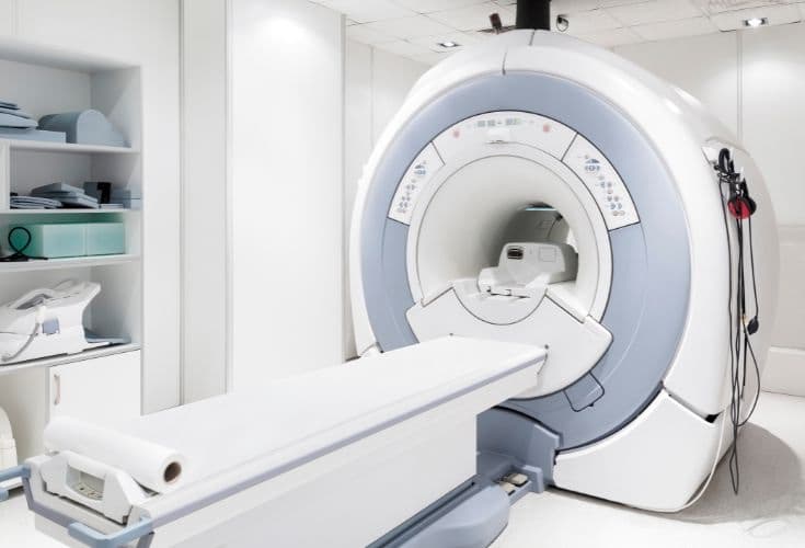
With modern technology, the 1.5 Tesla MRI system delivers images with high sharpness and can, in particular, produce 3D reconstructions of the scanned images, supporting image review and the accurate detection of morphological and structural lesions at an early stage, such as in blood vessels, the brain, the spine, and the head, face, and neck.…. helping physicians detect abnormalities accurately and quickly from the earliest stage of disease.
1.5 Tesla MRI Scanner System
- Model: 781341
- Manufacturer: Philips - Netherlands
Outstanding advantages of the 1.5 Tesla MRI system
- Modern, safe, and fast technology. Accurate results. Non-invasive and radiation-free. No contraindications for pregnant women and young children.
- Reduced noise during scanning.
- Sharp, detailed images. Detects even small lesions in hard-to-reach locations. Gives physicians a comprehensive view so that even the smallest lesions in the organs are not missed.
- Fast scan times. A whole-body scan takes just…seconds.
- Widely used to detect conditions of the brain, neck, spine, musculoskeletal system, cardiovascular system, oncology…
Medical applications
The 1.5 Tesla digital magnetic resonance imaging (MRI) scanner system is an important tool in medical imaging and is widely used in many different applications. The main medical applications of the 1.5 Tesla MRI scanner:
Diagnosis and monitoring of brain conditions
Used to diagnose and monitor brain problems, including brain nodular tumors, migraines, cerebrovascular conditions, and other conditions.
Imaging of bones, joints, and muscles
Evaluates problems of the bones, cartilage, muscles, and other related parts such as joints and vascular tissue.
Spine and pigmentation studies
Used to study and diagnose spine-related problems, especially back pain and pigmentation problems.
Imaging of the liver and abdominal organs
Evaluates problems of the liver, abdominal organs, and intestinal tract, helping diagnose liver tumors, liver pimples, and other conditions.
Imaging of the heart and blood vessels
Evaluates the structure and function of the heart and blood vessels, helping diagnose problems such as heart valve disease, cardiac nodular tumors, and vascular conditions.
Evaluation of chest conditions
Used to diagnose problems such as chest tumors, heart muscle conditions, and other chest-related problems.
Whole-body imaging
Evaluates the entire body to detect and monitor latent conditions and overall health.
Support for surgical decisions
Provides detailed information about the internal structures of the body, helping physicians plan and perform surgeries precisely.
Monitoring treatment effectiveness
Used to monitor the effectiveness of treatment methods, especially in cancer treatment and other conditions.
Medical research
Used in medical research to better understand the structure and function of the body, as well as to develop and evaluate new methods.
The 1.5 Tesla MRI scanner plays an important role in diagnosing and managing disease, providing high-quality images and detailed information to the medical team.
How should you prepare before an MRI scan?
Patients scheduled for an MRI of the abdomen, pelvis, or whole body need to fast for 4 hours before the scan.
Patients should inform the physician or the imaging technician if they have any devices or implants in their body, such as:
- Artificial heart valves
- Automatic drug infusion devices implanted under the skin
- Intrauterine devices (IUDs)
- Artificial joints or femoral heads
- Vascular clips or bone fixation plates and screws
For other cases, medical staff will provide instructions.
What should you keep in mind during an MRI scan?
- An MRI scan takes 15 to 45 minutes, depending on each specific case.
- Depending on the body part, the technician will give specific notes and instructions so that the images are clear and accurate.
- During the MRI scan, the patient stays alone in the scanner room, while the technician and the diagnostic imaging physician monitor through a glass window and a computer system connected to the scanner.
- When a contrast agent injection is needed, the patient may feel warmth throughout the body or a bitter taste on the tongue during the injection; this is normal, and the symptoms resolve on their own within 2 to 5 minutes.
HIU Hospital, with its modern facilities and medical equipment and a team of experts and physicians with many years of experience in examination and treatment, is a place where patients can feel completely at ease receiving examination and treatment.