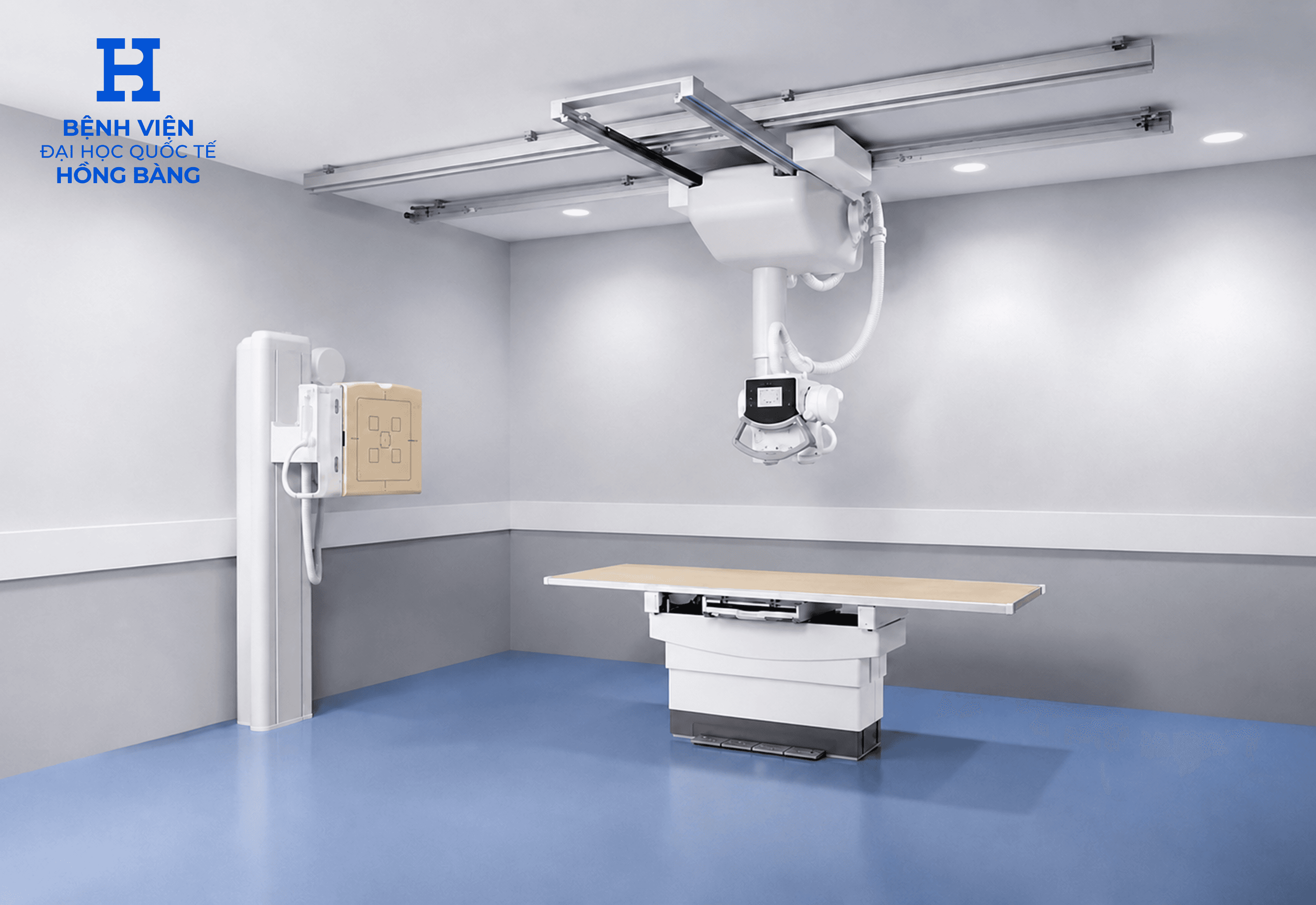
Ceiling-Mounted Digital X-ray System
- Model: Digital Diagnost
- Manufacturer: Philips
Key advantages of the ceiling-mounted digital X-ray system
- The UNIQUE (UNified Image Quality Enhancement) image post-processing software delivers outstanding image quality and accurate diagnostic results.
- With its ceiling-mounted design and intelligent control system, the Digital Diagnost ceiling-mounted digital X-ray system can perform flexible, precise movements, capturing intuitive, vivid two-dimensional images for clinical diagnosis. At the same time, the system can easily image patients in many different positions, such as standing, sitting, or lying down, and can reach every organ in the body.
- Widely used for clinical diagnosis in departments such as emergency, orthopedics…
What is an X-ray?
In 1895, X-rays were first discovered by the German physics professor Wilhelm Conrad Roentgen. As short-wavelength electromagnetic waves able to pass through all matter, X-rays have been applied by scientists in many fields, especially in medicine with the creation of X-ray machines, Ct-scanners, PET- CT, …
With the development of science and technology, X-ray equipment has also been gradually improved and upgraded with many modern technologies, including the Digital Diagnost ceiling-mounted digital X-ray system, which provides strong support in diagnostic imaging as well as in the treatment of many medical conditions. X-ray imaging is a simple, fast diagnostic imaging method with high diagnostic effectiveness. The X-ray machine emits a beam of X-rays that passes through the body area being imaged. In areas of soft tissue, fluid, or gas (lungs), more X-rays pass through, and the image on the film appears black. Dense body parts such as bones, muscles, or tumors block some of the X-rays, and the image of these areas appears gray or whiter, depending on the density of these body areas.
The human eye cannot see X-rays, and during an X-ray, the person being imaged feels no pain or discomfort.
Applications of the ceiling-mounted digital X-ray system
- Detects fractured bones and teeth, traumatic defects, or other abnormalities of the skeletal and dental system…
- Detects degenerative joint disease, arthritis, joint dislocation…
- Pleural or intestinal effusion and fluid collection…
- Detects tumors or certain abnormalities of the heart….
Are X-rays harmful?
X-ray imaging is a common diagnostic imaging technique today that helps physicians reach diagnoses quickly and effectively at low cost. However, many people also fear that X-rays may cause unwanted side effects, even radiation exposure or cancer. According to some studies, this concern is well-founded if a person is exposed to X-rays continuously over a long period.
Therefore, to ensure patients' health and safety, specialist physicians always examine patients in person and order X-rays appropriate to each specific condition. During imaging, the X-ray dose is also set to the minimum level that still meets film quality requirements while ensuring maximum safety for the patient.
Pregnant women in particular are advised not to have X-rays unless truly necessary, because X-rays can cause abnormalities in the fetus, especially in the early stage of pregnancy.
However, in practice, the risks that X-rays pose to human health are very low compared with the outstanding advantages that X-ray imaging brings to diagnostic imaging.
The Diagnostics Imaging Department of HIU Hospital is equipped with many modern, high-tech systems such as the Digital Diagnost ceiling-mounted digital X-ray system, along with a team of experienced, dedicated experts, diagnostic imaging physicians, and technicians, helping deliver accurate, fast diagnostic results to patients while saving time and cost.